AI in Medicine · Marie Skłodowska-Curie Fellow

Amir Reza Naderi Yaghouti

I develop machine learning and deep learning methods for medical data, from MRI, retinal and ultrasound images to clinical and laboratory records, to make diagnosis earlier and more reliable.

Citations
63
h-index
3
i10-index
2
Papers
4
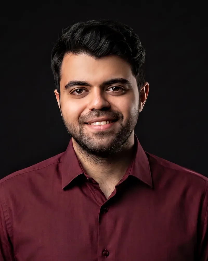
Portrait of Amir Reza Naderi Yaghouti

About

I'm Amir Reza Naderi Yaghouti, or Amir Naderi for short: a researcher in AI in medicine and a Marie Skłodowska-Curie Fellow in the EGRET-AAA network, doing a joint PhD at Otto-von-Guericke University Magdeburg and the University of Groningen.

My research applies AI across medicine: imaging, clinical records and laboratory data. In my PhD I use deep learning, foundation models and explainable AI to find signs of glaucoma in retinal images and brain MRI.

Before that, I built models for liver disease and ovarian cancer from ultrasound images, blood tests and routine lab biomarkers. I was born and raised in Tehran, where I studied Biomedical Engineering.

Position
Doctoral Candidate, EGRET-AAA (DC9)
Supervisors
Prof. Michael B. Hoffmann · Dr. Khaldoon O. Al-Nosairy · Prof. Nomdo M. Jansonius
Funding
EU Horizon Europe, MSCA grant 101072435
Languages
Persian (native) · English (C1) · German (A1)
Research

Ophthalmology · PhD

Glaucoma signs beyond the optic disc

Masking anatomical regions in fundus images shows which parts a retinal foundation model relies on to detect glaucoma, including vascular signal outside the optic disc.

  • Fundus images
  • Foundation models
  • Explainable AI

Neuroimaging · PhD

Optic chiasm degeneration on MRI

Deep learning segmentation of anatomical MRI measures how glaucoma affects the optic chiasm, where the visual pathways cross in the brain.

  • MRI
  • Segmentation

Hepatology

Fatty liver disease and NASH

Grading fatty liver on B-mode ultrasound with pre-trained CNNs, and detecting non-alcoholic steatohepatitis early from clinical and blood parameters.

  • Ultrasound
  • Clinical data
  • Radiomics

Oncology

Ovarian cancer detection

A review of a decade of AI for ovarian cancer imaging, and a tabular foundation model that separates benign from malignant tumors using routine lab biomarkers.

  • Lab biomarkers
  • Medical imaging
  • Foundation models

Publications

Synced daily from Google Scholar

All on Google Scholar
Area
Task
Method
Data

Showing 5 of 5

  1. Sci. Rep. · under review

    Discrimination of benign and malignant ovarian tumors from routine laboratory biomarkers using a tabular foundation model

    AR Naderi Yaghouti, A Shalbaf

    • Ovarian cancer
    • Classification
    • Deep learning
    • Foundation models
    • Clinical & lab data
  2. J. Biomed. Phys. Eng.2025

    Classification of Nonalcoholic Fatty Liver Grades using Pre-Trained Convolutional Neural Networks and a Random Forest Classifier on B-Mode Ultrasound Images

    AR Naderi Yaghouti, A Shalbaf

    Journal of Biomedical Physics and Engineering

    • Liver disease
    • Classification
    • Machine learning
    • Deep learning
    • Ultrasound
    • Medical imaging
    PaperScholar
    Cite
    @article{naderiyaghouti2025classification,
      title = {{Classification of Nonalcoholic Fatty Liver Grades using Pre-Trained Convolutional Neural Networks and a Random Forest Classifier on B-Mode Ultrasound Images}},
      author = {Naderi Yaghouti, A. R. and Shalbaf, A.},
      journal = {Journal of Biomedical Physics and Engineering},
      year = {2025}
    }
    7 citations
  3. Arch. Comput. Methods Eng.2025

    Artificial intelligence for ovarian cancer detection with medical images: a review of the last decade (2013–2023)

    AR Naderi Yaghouti, A Shalbaf, R Alizadehsani, RS Tan, A Vijayananthan, ...

    Archives of Computational Methods in Engineering 32 (7), 4093-4124

    • Ovarian cancer
    • Review
    • Medical imaging
    PaperDOIScholar
    Cite
    @article{naderiyaghouti2025artificial,
      title = {{Artificial intelligence for ovarian cancer detection with medical images: a review of the last decade (2013–2023)}},
      author = {Naderi Yaghouti, A. R. and Shalbaf, A. and Alizadehsani, R. and Tan, R. S. and Vijayananthan, A. and others},
      journal = {Archives of Computational Methods in Engineering},
      volume = {32},
      number = {7},
      pages = {4093--4124},
      year = {2025},
      doi = {10.1007/s11831-025-10268-x}
    }
    15 citations
  4. Sci. Rep.2024

    Machine learning approaches for early detection of non-alcoholic steatohepatitis based on clinical and blood parameters

    AR Naderi Yaghouti, H Zamanian, A Shalbaf

    Scientific Reports 14, 2442

    • Liver disease
    • Classification
    • Machine learning
    • Clinical & lab data
    PaperDOIScholar
    Cite
    @article{naderiyaghouti2024machine,
      title = {{Machine learning approaches for early detection of non-alcoholic steatohepatitis based on clinical and blood parameters}},
      author = {Naderi Yaghouti, A. R. and Zamanian, H. and Shalbaf, A.},
      journal = {Scientific Reports},
      volume = {14},
      pages = {2442},
      year = {2024},
      doi = {10.1038/s41598-024-51741-0}
    }
    38 citations
  5. Tehran Univ. Med. J.2021

    Automatic classification of Non-alcoholic fatty liver using texture features from ultrasound images

    AR Naderi Yaghouti, A Shalbaf, A Maghsoudi

    Tehran University Medical Journal 79 (1), 10–17

    • Liver disease
    • Classification
    • Machine learning
    • Ultrasound
    • Medical imaging
    PaperScholar
    Cite
    @article{naderiyaghouti2021automatic,
      title = {{Automatic classification of Non-alcoholic fatty liver using texture features from ultrasound images}},
      author = {Naderi Yaghouti, A. R. and Shalbaf, A. and Maghsoudi, A.},
      journal = {Tehran University Medical Journal},
      volume = {79},
      number = {1},
      pages = {10--17},
      year = {2021}
    }
    3 citations

News

  1. TalkOral presentation at CLINICCAI 2026 (MICCAI 2026 Clinical Day) in Strasbourg, France: “Glaucoma Biomarkers Beyond the Optic Disc: Vascular Signal Quantified via Anatomical Masking of a Retinal Foundation Model”. Read more (opens in new tab)

  2. PosterPoster presentation at EVER 2026, the European Association for Vision and Eye Research conference, in Florence, Italy.

  3. PosterPoster presentation at ECVP 2025, the European Conference on Visual Perception, in Mainz, Germany.

  4. PaperNew review in Archives of Computational Methods in Engineering: artificial intelligence for ovarian cancer detection with medical images over the last decade (2013–2023). Read more (opens in new tab)

  5. PaperNew paper in the Journal of Biomedical Physics and Engineering: classifying nonalcoholic fatty liver grades with pre-trained CNNs and a random forest on B-mode ultrasound. Read more (opens in new tab)

  6. MilestoneStarted a joint PhD in Visual Neuroscience at Otto-von-Guericke University Magdeburg and the University of Groningen as a Marie Skłodowska-Curie Doctoral Candidate (EGRET-AAA, Project 9), working on deep learning for visual pathway anomalies in glaucoma. Read more (opens in new tab)

Show earlier news
  1. PaperNew paper in Scientific Reports: machine learning approaches for early detection of non-alcoholic steatohepatitis (NASH) from clinical and blood parameters. Read more (opens in new tab)

Talks

Oral · 2026

CLINICCAI

MICCAI Clinical Day · Strasbourg, France

Glaucoma Biomarkers Beyond the Optic Disc: Vascular Signal Quantified via Anatomical Masking of a Retinal Foundation Model
Poster · 2026

EVER

European Association for Vision and Eye Research · Florence, Italy

Poster · 2025

ECVP

European Conference on Visual Perception · Mainz, Germany

Education

Jun 2024 – 2027 (expected) · Joint doctorate

PhD, Visual Neuroscience

Otto-von-Guericke University Magdeburg · University of Groningen / UMC Groningen

Marie Skłodowska-Curie Doctoral Fellow, EGRET-AAA

Sep 2018 – Oct 2021 · GPA 18.43 / 20 (3.77 / 4.0)

MSc, Biomedical Engineering

Islamic Azad University, Science and Research Branch, Tehran

Thesis: Transfer learning with deep convolutional neural networks for classification of liver steatosis: a comparison of radiomics and convolutional features

Sep 2014 – Jul 2018 · Final-year GPA 18.09 / 20

BSc, Biomedical Engineering

Islamic Azad University, Science and Research Branch, Tehran

Thesis: Automated breast lesion segmentation and classification in ultrasound imaging via texture analysis

Honors

2024

Marie Skłodowska-Curie Doctoral Fellowship, EU Horizon Europe

2021

Highest possible grade for the Master's thesis

2021

Top student of the Master's program, with the highest grades in most courses

Toolkit

Methods
Deep learning · foundation models · segmentation · explainable AI · radiomics · machine learning on clinical data · signal processing · NLP
Data
MRI · fundus photography · ultrasound · clinical records · laboratory biomarkers
Code
Python (PyTorch, TensorFlow, MONAI, scikit-learn, OpenCV) · MATLAB · R
Tools
LaTeX · Git · SPSS · Minitab · RapidMiner

Contact

Let's talk about AI in medicine.

Open to research collaborations in medical AI, from imaging to clinical data.

Personal email: naderi.bme@gmail.comDownload CV (PDF)